Thymus
A gland behind the breastbone where T cells, a kind of white blood cell, mature.
Thymus anatomy is that of an organ of the immune system where T cells, white blood cells of the adaptive immune response, mature. With the bone marrow it is one of the two primary lymphoid organs, the places where lymphocytes mature and are selected so that they attack germs without harming the body's own cells. The thymus is relatively large in infants and children and quite small in older adults. Its slow shrinking, called involution, takes about 3 per cent of its tissue a year from birth to age 35–45, then about 1 per cent a year.
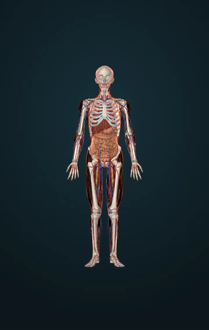
It lies in the upper chest, in the space between the breastbone in front and the aorta behind, above the heart. When it is growing most actively, it reaches from the fourth rib cartilage up to the lower border of the thyroid gland, partly in the chest and partly in the neck. The breastbone and the origins of two neck muscles, the sternohyoid and sternothyroid, cover it; below, it rests on the pericardium, the sac round the heart, separated from the arch of the aorta and the great vessels by a layer of fascia.
It has two lobes, usually of different sizes, held together and enclosed by connective tissue that forms its capsule. The capsule sends partitions, the trabeculae, inwards, dividing it into lobules. The outer part of each lobule, the cortex, is packed with developing T cells, called thymocytes, together with some epithelial cells, macrophages and dendritic cells. The inner medulla holds fewer thymocytes, epithelial and dendritic cells, and nest-like bodies, Hassall's corpuscles.
T cells start in the bone marrow; as thymocytes they move to the thymus and mature there. In the cortex, positive selection keeps those that can bind the body's own MHC molecules, the proteins that display antigens. At the junction of cortex and medulla, negative selection kills by apoptosis those that bind the body's own antigens. Only about 2 per cent of the thymocytes that enter the thymus leave it as mature T cells. They enter the blood and go to other lymphatic organs, where they help defend the body. The thymus also makes hormones, the thymosins, which help T cells develop.
Numbers
| Thymocytes leaving as mature T cells | about 2% | OpenStax, Anatomy and Physiology (1st ed., CC BY-NC-SA 4.0: linked, never copied) — 21.3 The Adaptive Immune Response: T lymphocytes and Their Functional Types |
|---|---|---|
| Extent when growing most actively | from the 4th rib cartilage up to the lower border of the thyroid gland | Gray, Anatomy of the Human Body (20th ed., 1918, public domain), The Thymus, pp. 1273–1274 |
Its parts in the model
- Left lobe of thymus
- Right lobe of thymus
Sources
- OpenStax, Anatomy and Physiology (1st ed., CC BY-NC-SA 4.0: linked, never copied) — 21.3 The Adaptive Immune Response: T lymphocytes and Their Functional Types
- Gray, Anatomy of the Human Body (20th ed., 1918, public domain), The Thymus, pp. 1273–1274
- OpenStax, Anatomy and Physiology (1st ed., CC BY-NC-SA 4.0: linked, never copied) — 21.1 Anatomy of the Lymphatic and Immune Systems
- OpenStax, Anatomy and Physiology (1st ed., CC BY-NC-SA 4.0: linked, never copied) — 17.10 Organs with Secondary Endocrine Functions
- NCI SEER Training Modules — Thymus
- BodyParts3D, © The Database Center for Life Science, CC BY-SA 2.1 Japan — adapted.