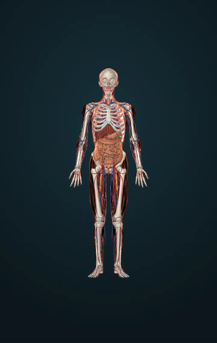
Urinary bladder
A stretchy muscular bag in the pelvis that stores urine until it leaves through the urethra.
Bladder anatomy is that of a temporary reservoir: a hollow, highly stretchy muscular organ whose size and shape change with the amount of urine it holds and with the pressure of the organs around it. Urine made in the kidneys reaches it down the two ureters, and it leaves through the urethra. In an adult the bladder holds anything from nearly nothing to 500–600 mL.
It sits in the pelvis between the hip bones, behind the pubic symphysis and in front of the rectum. In women the uterus lies behind it; in men the prostate lies just below it. It lies beneath the peritoneum, which covers only its top, the dome. As the bladder fills, the dome rises upward.
Its wall has four layers. The lining is transitional epithelium, the same as in the ureters: its cells look tall when the bladder is empty and flatten as it stretches, and the empty lining lies in folds called rugae. Beneath it is the submucosa, connective tissue with elastic fibres. Then comes the detrusor muscle, smooth muscle whose fibres cross in all directions. Outside, peritoneum covers the top and fibrous tissue the rest. On the floor, the two ureters' openings and the opening of the urethra mark out a triangle, the trigone. A band of the detrusor round the urethral opening forms the internal urethral sphincter.
Emptying the bladder, called micturition, rests on a spinal reflex. Stretch receptors in the wall send signals to the sacral part of the spinal cord. At about 150 mL a first urge is felt, which is easily put off; the urges grow harder to ignore, and at 300–400 mL holding on fails. While the bladder fills, sympathetic nerves hold the detrusor back, and the external urethral sphincter, skeletal muscle under conscious control through the pudendal nerve, stays shut. To empty, parasympathetic nerves make the detrusor contract and the internal sphincter relax, and the external sphincter lets go. Babies empty the bladder by this reflex; children learn to delay it by holding the external sphincter.
Numbers
| Capacity, adult | from nearly 0 to 500–600 mL | OpenStax, Anatomy and Physiology (1st ed., CC BY-NC-SA 4.0: linked, never copied) — 25.2 Gross Anatomy of Urine Transport |
|---|---|---|
| First urge to pass urine | at about 150 mL | OpenStax, Anatomy and Physiology (1st ed., CC BY-NC-SA 4.0: linked, never copied) — 25.2 Gross Anatomy of Urine Transport |
| Urine the kidneys make | about 1–2 quarts (1–2 litres) a day | NIDDK — The Urinary Tract & How It Works |
Its parts in the model
- Urethra
- Urinary bladder
Sources
- OpenStax, Anatomy and Physiology (1st ed., CC BY-NC-SA 4.0: linked, never copied) — 25.2 Gross Anatomy of Urine Transport
- NIDDK — The Urinary Tract & How It Works
- NCI SEER Training Modules — Urinary Bladder
- BodyParts3D, © The Database Center for Life Science, CC BY-SA 2.1 Japan — adapted.