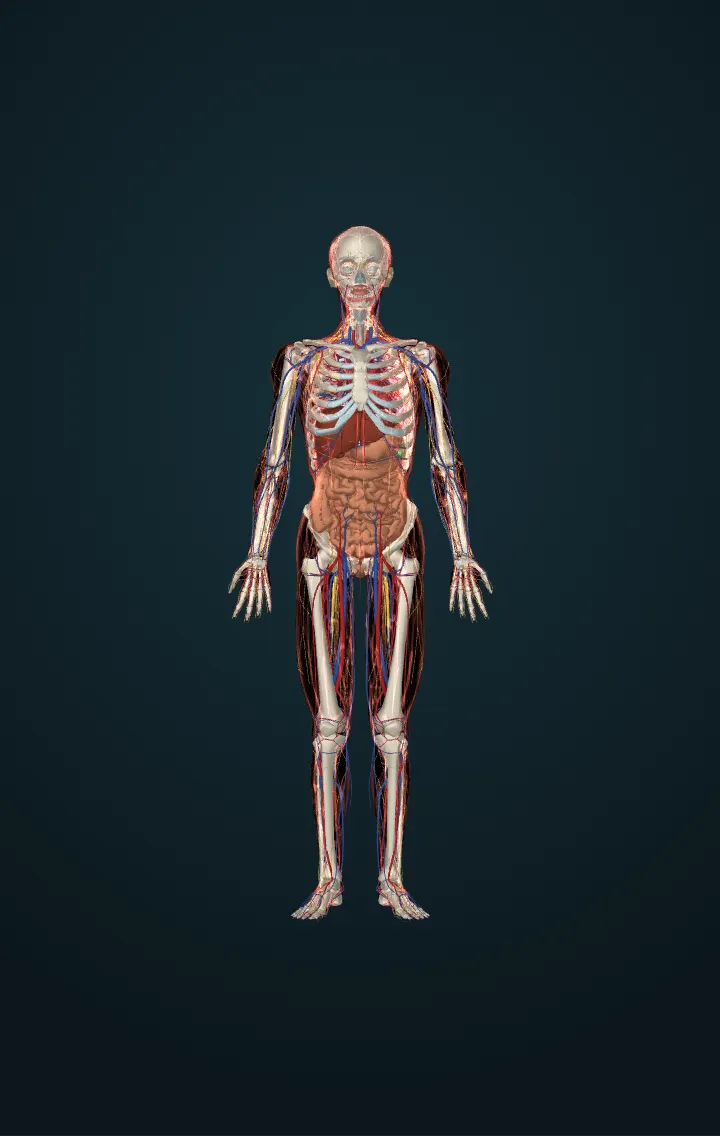
Heart
A muscular pump in the middle of the chest that drives blood through the lungs and the whole body.
The heart lies in the middle of the chest, between the lungs, with its tip turned to the left. It has four chambers: two atria that receive blood and two ventricles that pump it out. The right side sends blood to the lungs to pick up oxygen; the left side sends it to the rest of the body.
Four valves keep the blood moving one way. Each beat starts as an electrical signal in the sinoatrial node, spreads across the atria, pauses at the atrioventricular node and then sets off the ventricles. The sounds of a heartbeat are the valves closing.
Numbers
| Resting heart rate, adult | 60–100 beats a minute | MedlinePlus — Pulse |
|---|---|---|
| Blood pumped at rest | about 5 litres a minute | OpenStax, Anatomy and Physiology (1st ed., CC BY-NC-SA 4.0: linked, never copied) |
| Size | about the size of a fist | NHLBI — How the heart works |
Its parts in the model
- Anterior cardiac vein
- Anterior interventricular vein
- Anterior leaflet of mitral valve
- Anterior leaflet of tricuspid valve
- Anterior papillary muscle of right ventricle
- Anterior semilunar cusp of pulmonary valve
- Anterolateral head of lateral papillary muscle of left ventricle
- Ascending aorta
- Atrioventricular bundle
- Atrioventricular node
- Chordae tendineae of mitral valve
- Chordae tendineae of tricuspid valve
- Circumflex branch of left coronary artery
- Conus branch of anterior interventricular branch of left coronary artery
- Coronary sinus
- Diagonal branch of anterior descending branch of left coronary artery
- First anterior ventricular branch of right coronary artery
- First posterior ventricular branch of right coronary artery
- First right anterior branch of anterior interventricular branch of left coronary artery
- First septal branch of right posterior interventricular artery
- Free wall of left ventricle
- Free wall of right ventricle
- Great cardiac vein
- Interventricular septum
- Lateral papillary muscle of left ventricle
- Left bundle branch
- Left coronary cusp of aortic valve
- Left fibrous ring
- Left inferior pulmonary vein
- Left marginal vein
- Left semilunar cusp of pulmonary valve
- Left superior pulmonary vein
- Marginal branch of right coronary artery
- Middle cardiac vein
- Non-coronary cusp of aortic valve
- Posterior interventricular branch of right coronary artery
- Posterior leaflet of mitral valve
- Posterior leaflet of tricuspid valve
- Posterior papillary muscle of right ventricle
- Posterior vein of left ventricle
- Pulmonary trunk
- Right bundle branch
- Right conus artery
- Right coronary cusp of aortic valve
- Right fibrous ring
- Right inferior pulmonary vein
- Right marginal vein
- Right semilunar cusp of pulmonary valve
- Right superior pulmonary vein
- Second right anterior branch of anterior interventricular branch of left coronary artery
- Second septal branch of right posterior interventricular artery
- Septal branch of anterior interventricular artery
- Septal leaflet of tricuspid valve
- Septal papillary muscle of right ventricle
- Septomarginal trabecula
- Sinuatrial node
- Small cardiac vein
- Subendocardial branches
- Superior vena cava
- Third right anterior branch of anterior interventricular branch of left coronary artery
- Trunk of anterior interventricular branch of left coronary artery
- Trunk of left coronary artery
- Trunk of right coronary artery
- Wall of left atrium
- Wall of right atrium
Sources
- MedlinePlus — Pulse
- OpenStax, Anatomy and Physiology (1st ed., CC BY-NC-SA 4.0: linked, never copied)
- NHLBI — How the heart works
- BodyParts3D, © The Database Center for Life Science, CC BY-SA 2.1 Japan — adapted.